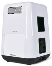
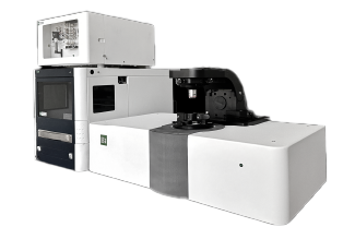
VISQUE InVivo Smart-LF – パーソナル in vivo 発光・蛍光・近赤外蛍光イメージングシステム (Vieworks)
Visque Smart-LFの特長
- 高感度冷却sCMOSカメラを搭載
- 可視~近赤外領域の蛍光・発光検出(500nm – 860 nm)
- 5波長のLED励起光源を標準搭載(Blue | Green | Red | HyperRed | NIR)
- 卓上型のコンパクトサイズ(本体:40✕40✕57 cm, 重量:22kg) ※別途PC,モニタ付属
- 最大でマウス3匹の同時計測が可能
- 最大30フレーム/秒以上のタイムラプス計測が可能
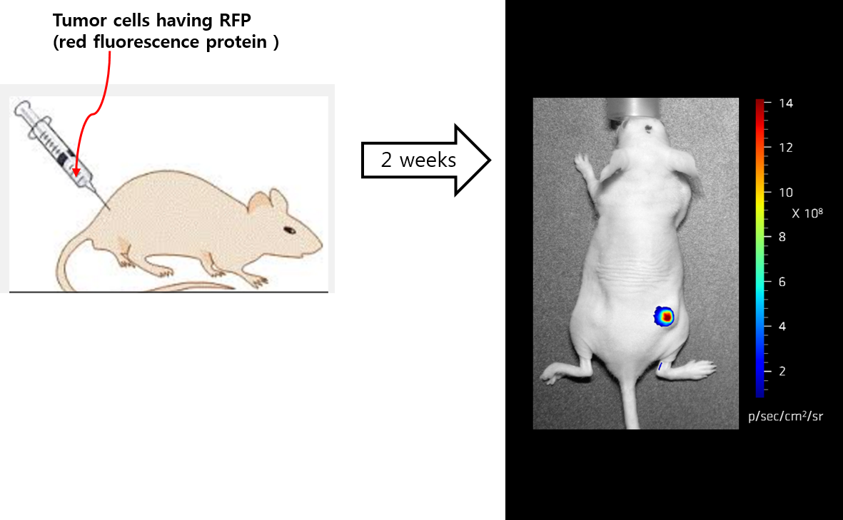
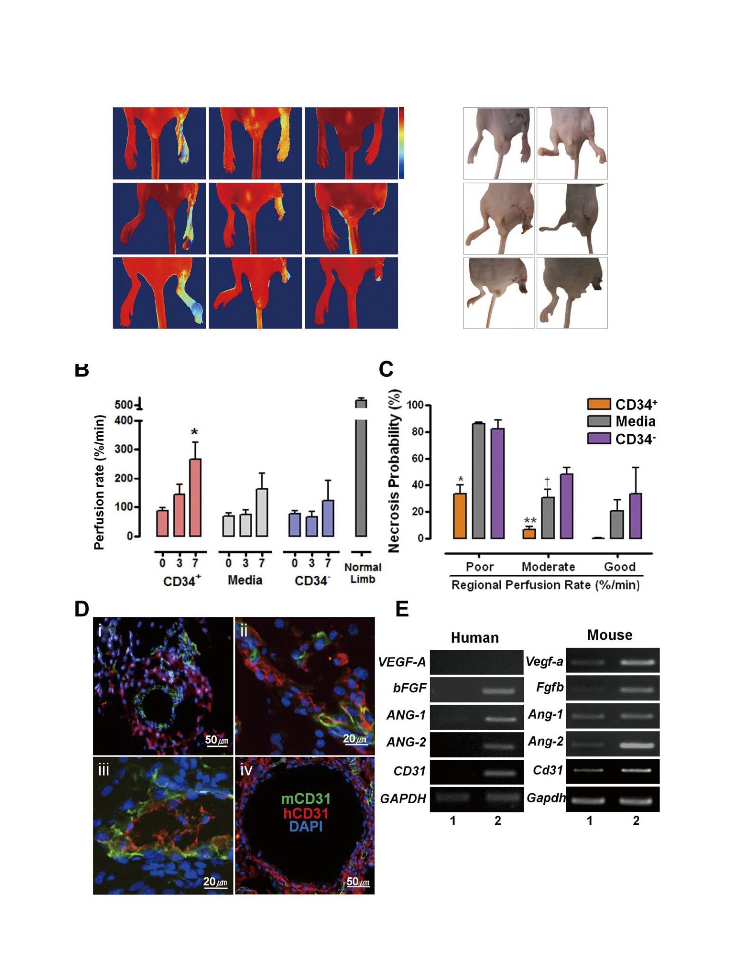
蛍光イメージング
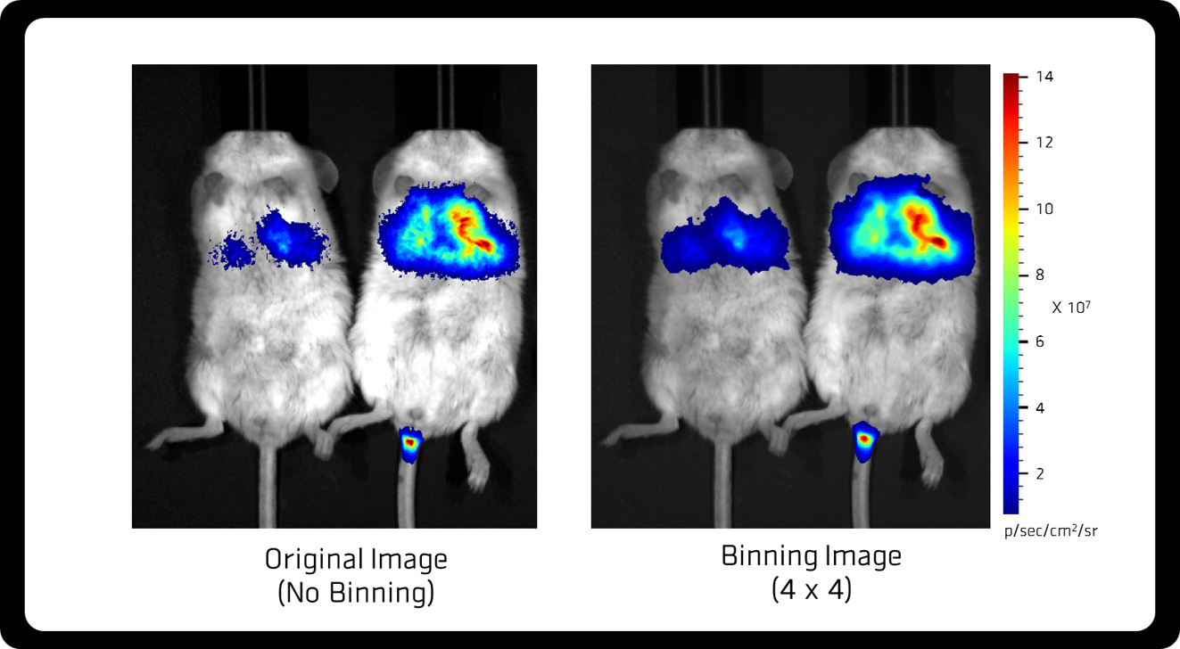
発光イメージング
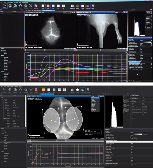
タイムラプス
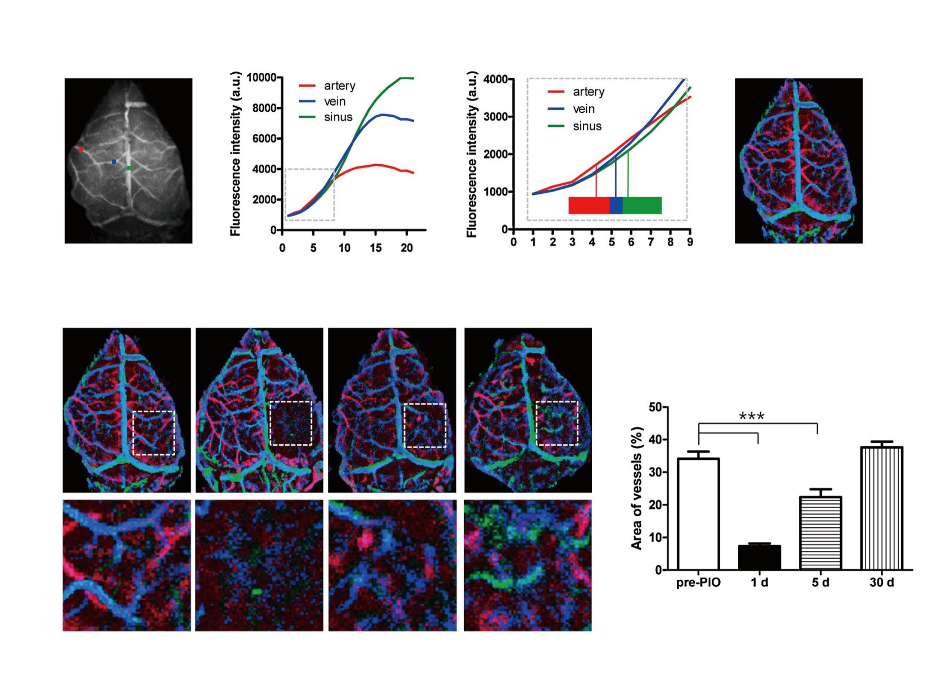
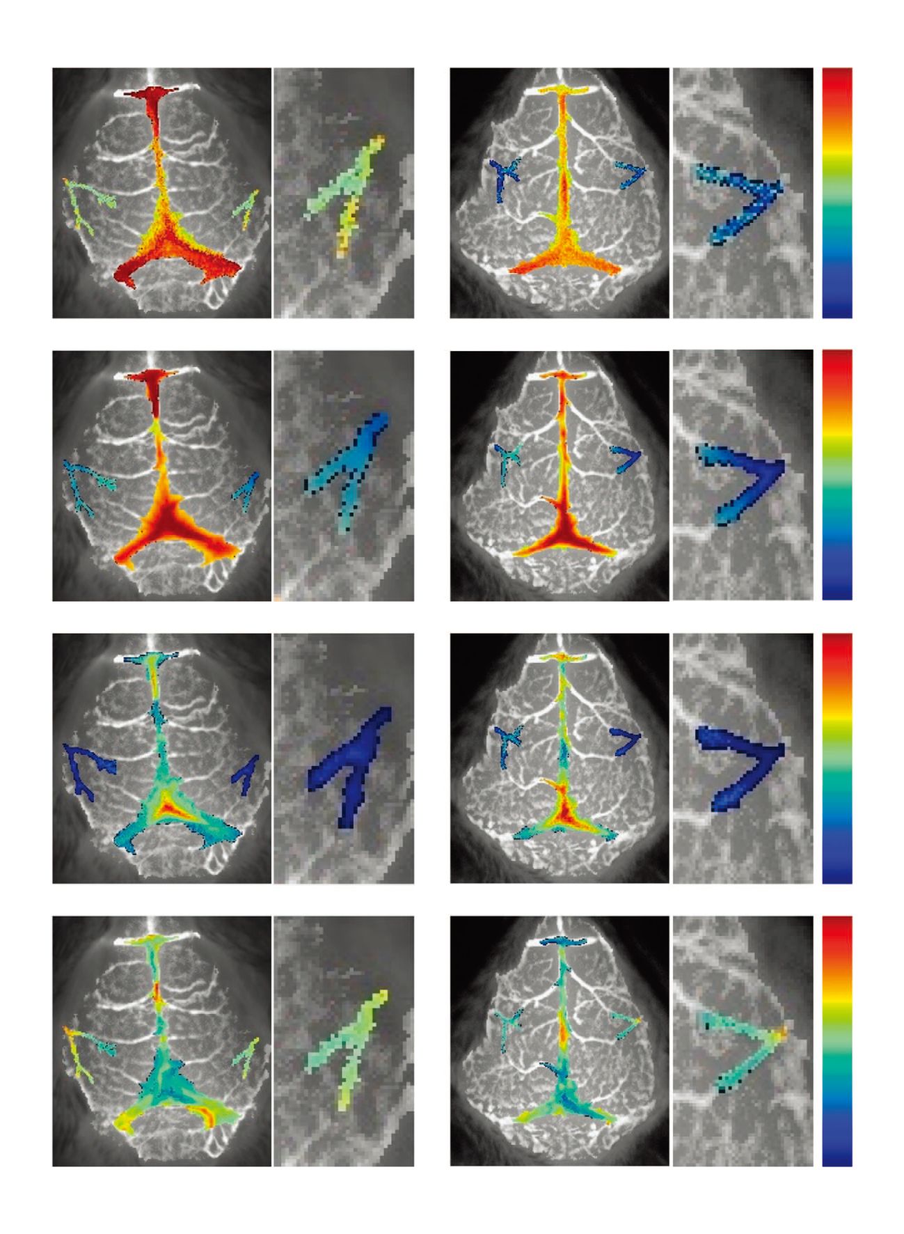
マウス脳のリアルタイム画像の比較 組織灌流 マウス 月齢2ヶおよび12ヶ月のマウス
「マウスの骨格構造および血流の年齢に関連した変化」
“Age-related changes in pialarterial structure and blood flow in mice”
NeurobiolAging. 2015 sept 11 (Accepted)
下のタイムラプス画像は尾部の静脈に近赤外線の蛍光色素であるICGを注入した後、脳の骨の頭部の皮膚を開き、脳血管のICG循環をVisqueで撮影。 計100枚の画像を1秒間に約6回撮影。 左の画像は月齢2ヶ月のマウスの血流を示し、右の画像は月齢12ヶ月のマウスの血流を示しています。 この画像は慶煕大学の解剖学科により経年変化による血管および血流変化の研究として発表されました。
※NeurobiolAging, SCI(Science Citation Indexに基づくImpact Factor 5.1。
ソフトウェア
VISQUEには薬物動態/ファーマコキネティクス解析および生体内分布解析のための10種類以上のアルゴリズムをサポートするソフトウェア「CleVueTM」が付属しています。CleVueTMには以下の特長があります。
- 4種類の違う画像を比較して同時に解析可能
- 最適のウィンドウ幅で自動調整
- 各ピクセルのカイネティックカーブはタイムラプス画像で利用
- 計測・解析操作を容易に実施できる直観的なインターフェース
- 計測データの解析・編集がしやすいCIF形式のデータ出力
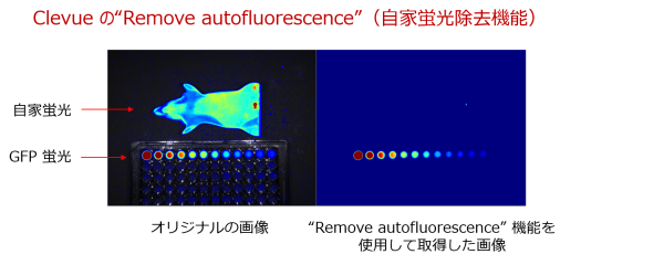
Clevueの自家蛍光除去機能
Clevueは自家蛍光を除去する機能を持っています。以下の画像例では、Clevueによる自家蛍光除去機能を使用して撮影した例になります。左の画像は元の画像を示しています。ファントムマウスに示されている蛍光はマウスの自家蛍光です。下の円の蛍光シグナルは、異なる濃度の緑色蛍光色素を希釈することにより蛍光シグナルの強度がマウスの自家蛍光の同様のレベルに調整された緑色蛍光色素を示しています。Clevueの自家蛍光を除去する機能を使用すれば自動で自家蛍光を取り除き目的の蛍光シグナルのみを捉えることができます。この機能は緑色と赤色の色の範囲を持つ蛍光のシグナル画像に適用することができます。
スペック情報
システム
| 装置名 | VISQUE™ InVivo Smart-LF |
| 測定項目 | In Vivo イメージング、発光・蛍光・近赤外・リアルタイムイメージング |
カメラ
| 画像センサー | 1.2″ バックサイドイルミネーションsCMOS |
| 冷却 | ⊿-20℃ (室温から-20℃冷却) ペルチェ素子 |
| 分解能 (H ✕ V) | 1824 ✕ 1824 |
| ピクセルサイズ | 6.5 μm ✕ 6.5 μm |
| 露光時間 | 25 ms – 15 min |
| 最大フレーム数 | 37 フレーム / 秒 |
| デジタル出力 | 16 bit |
| ビニング | 1 ✕ 1, 2 ✕ 2, 4 ✕ 4 |
| 蛍光検出: 光源 | LED |
| 蛍光検出:蛍光フィルター | 最大9枚(標準で4枚搭載) |
レンズ
| コントロール | モーター駆動(絞り、ズーム、フォーカス) |
| ズーム(視野、H ✕ V) | 15 cm ✕ 15 cm (1 x) – 5 cm ✕ 5cm (3 x) |
CleVue™ ソフトウェア
| 画像取得モード | シングルフレーム、アキュミレーション、タイムラプス |
| 画像形式 | cif, tif, bmp, jpg, png |
| カイネティクス解析 | 動力学グラフの作成、10種類のカイネティクス解析アルゴリズムを搭載 |
| 画像解析 | 自家蛍光除去、マルチ蛍光測定イメージの統合 |
ステージ
| ステージタイプ | スライドタイプのステージ、最大マウス3匹 |
| その他 | 加温機能、麻酔器用アダプター |
検出可能な蛍光波長(標準仕様)
| 励起光 | 励起波長(nm) | 検出フィルター | 検出波長(nm) | 蛍光色素 |
|---|---|---|---|---|
| Blue | 390 – 490 | GFP | 500 – 550 | GFP / EGFP / Alexa 488 / FITC / QD 525 |
| Green | 530 – 570 | PE | 575 – 640 | RFP / DsRed / PE / Alexa 568 / TRITC / QD 585 / QD 605 / QD 625 |
| Red | 620 – 650 | Cy 5.5 | 690 – 740 | Cy5.5 / PKE 680 / Alexa 680 / Alexa 700 / QD 705 |
| HyperRed | 630 – 680 | Cy 5.5 | 690 – 740 | Cy5.5 / PKE 680 / Alexa 680 / Alexa 700 / QD 705 |
| NIR | 740 – 790 | ICG | 810 – 860 | ICG / QD 800 |
カタログ
文献
- Optical measurement of mouse differences in cerebral blood flow using indocyanine green. JCBFM 2015
- Exendin-4 protects hindlimb ischemic injury by inducing angiogenesis. BBRC 2015
- Application of dynamic indocyanine green perfusion imaging for evaluation of vasoactive effect of acupuncture: a preliminary follow-up study on normal healthy volunteers. MDER 2014
- Noninvasive optical measurement of cerebral blood flow in mice using molecular dynamics analysis of indocyanine green. PLoS One 2012
- Segmental analysis of indocyanine green pharmacokinetics for the reliable diagnosis of functional vascular insufficiency. JBO 2011
- Dynamic fluorescence imaging for multiparametric measurement of tumor vasculature. JBO 2011
- Efficient differentiation of human pluripotent stem cells into functional CD34+ progenitor cells by combined modulation of the MEK/ERK and BMP4 signaling pathways. blood 2010
- Assessment of peripheral tissue perfusion by optical dynamic fluorescence imaging and nonlinear regression modeling. Proc of SPIE 2010
- Unsorted human adipose tissue-derived stem cells promote angiogenesis and myogenesis in murine ischemic hindlimb model. MicroVasc Res 2010
- Dynamic fluorescence imaging of indocyanine green for reliable and sensitive diagnosis of peripheral vascular insufficiency. Micro Vasc Res 2010
- Application of novel dynamic optical imaging for evaluation of peripheral tissue perfusion. IJC 2010
- Quantitative analysis of peripheral tissue perfusion using spatiotemporal molecular dynamics. Plos One 2009
